1. 3D cell culture substrate made of fungus-derived cellulose nanofibers
Download the product flyer >>
Download the Product Data Sheet >>
Download the Safety Data Sheet >>
Three-dimensional (3D) culture of cells was realized using nanofiber cellulose synthesized by acetic acid bacteria from sugar beet (sugar beet) in Hokkaido, and we succeeded in producing spheroids of various cells (Example 1-1). For example, HepG2 spheroids with high activity of drug-metabolizing enzymes (CYP molecular species) inherent in the liver are produced by three-dimensional culture of HepG2 hepatocellular carcinoma cells (Example 1-2), which enables us to conduct high-throughput drug metabolism studies. In addition, iPS spheroids can be generated by three-dimensional culture of iPS cells without any special manipulation, which maintains undifferentiated growth (Example 1-3).
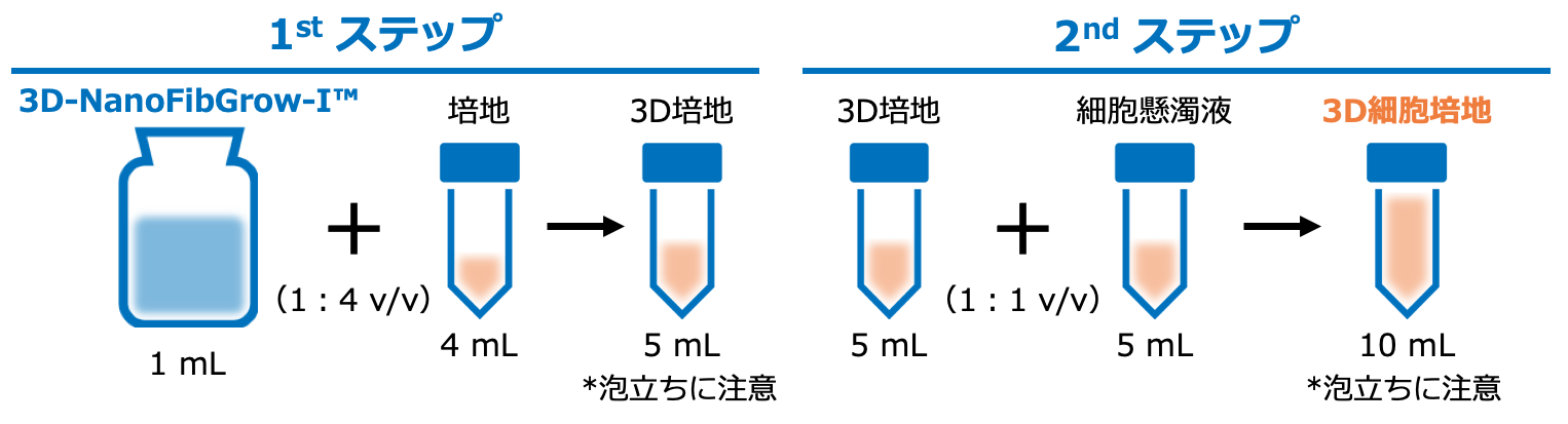
Instructions for use (for the preparation of 10 mL of 3D cell medium)
*The 3D culture at the interface of the wells with the product on the bottom of the wells (stacking method) is also available. Please contact us for details.
特徴
- Preparation of 3D cell culture media in two easy steps.
- Depending on the application, you can choose from a variety of culture vessels (well plate, flask, dish, etc.) and simply add 3D cell culture media to your chosen culture vessel to achieve 3D cell culture.
- No other special reagents or equipment is required.
- Sterilized, so there’s no need to worry about contamination.
- Operates at room temperature, so no complicated temperature control is required.
- Various media (validated reference media: RPMI-1640, D-MEM)
- Various media (validated reference media: RPMI-1640, D-MEM)
Recommended cell concentration: 1.0 x 10⁴cells/mL 〜 1.0 × 10⁶cells/mL
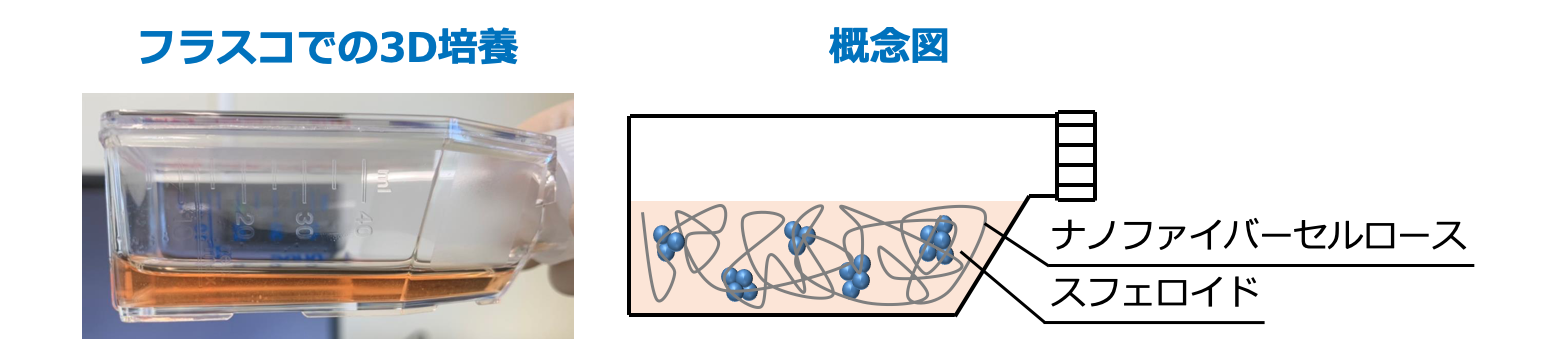
3D culture image
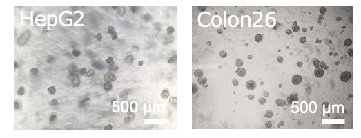
Example 1-1: Cell lines that can be cultured in 3D
Colon26 mouse colon cancer cells, HepG2 human liver cancer cells, MCF-7 human breast cancer cells, 4T1 mouse breast cancer cells, MKN45 human stomach cancer cells, B16 mouse melanoma cells, and human iPS cells.
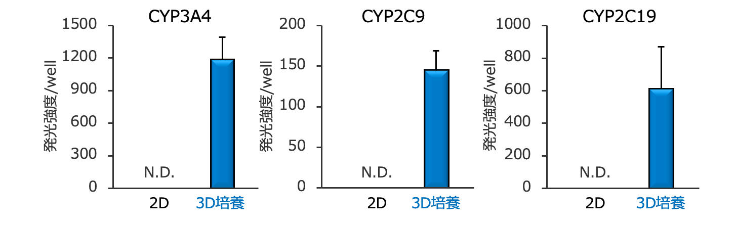
Example 1-2: Evaluation of the Activity of HepG2 Spheroid Drug Metabolizing Enzyme (CYP Molecular Species)
We have shown that 3D culture of HepG2 human liver cancer cells increases the activity of several CYP molecular species, which will allow evaluation of drug metabolism without the use of animals.